SPINE PAGE
-
Nomenclature and Classification of Lumbar Disc Pathology
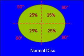
- Normal disc
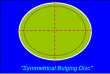
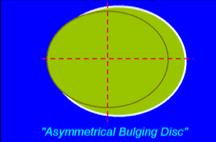
- Bulging discA disc in which the contour of the outer anulus extends, or appears to extend, in the horizontal (axial) plane beyond the edges of the disc space, over greater than 50% (180 degrees) of the circumference of the disc and usually less than 3mm beyond the edges of the vertebral body apophyses.
- Radial fissure or tear(B): Disruption of annular fibers extending from the nucleus outward toward the periphery of the annulus, usually in the vertical (cranio-caudal) plane, with occasional horizontal (transverse) components.
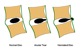
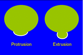
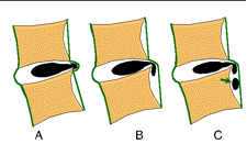
- ProtrusionProtrusion is a herniated disc in which the greatest distance, in any plane, between the edges of the disc material beyond the disc space is less than the distance between the edges of the base in the same plane. The test of protrusion is that there must be a localized (less than 50% or 180 degrees of the circumference of the disc.
Focal protrusion: Protrusion of disc material so that the base of the displaced material is less than 25% (90 degrees) of the circumference of the disc.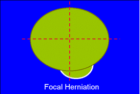
Broad-based protrusion: Herniation of disc material extending beyond the outer edges of the vertebral body apophyses over an area greater than 25% (90 degrees) and less than 50% (180 degrees) of the circumference of the disc.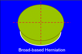
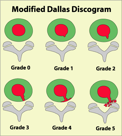
- ExtrutionA herniated disc in which, in at least one plane, any one distance between the edges of the disc material beyond the disc space is greater than the distance between the edges of the base in the same plane, or when no continuity exists between the disc material beyond the disc space and that within the disc space.
- Sequestration(C): An extruded disc in which a portion of the disc tissue is displaced beyond the outer annulus and maintains no connection by disc tissue with the disc of origin.
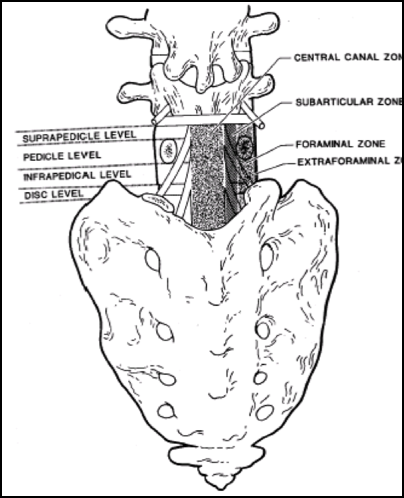
- ZonesAnterior zone: Peridiscal zone that is anterior to the mid-coronal plane of the vertebral body.
Central zone: Zone within the vertebral canal between sagittal planes through the medial edges of each facet.
Sub-articular zone: The zone, within the vertebral canal, sagittally between the plane of the medial edges of the pedicles and the plane of the medial edges of the facets, and coronally between the planes of the posterior surfaces of the vertebral bodies and the under anterior surfaces of the superior facets.
Foraminal zone: The zone between planes passing through the medial and lateral edges of the pedicles.
Extra-foraminal zone: The zone beyond the sagittal plane of the lateral edges of the pedicles, having no well-defined lateral border.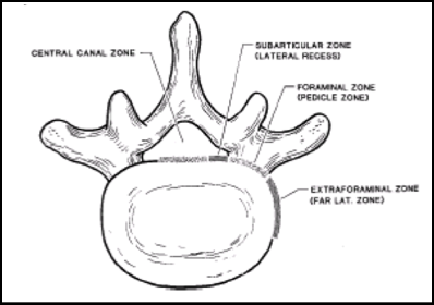
- LevelsDisc level: The level of the intervertebral disc.
Supra-pedicular level: The level within the vertebral canal between axial planes of the superior end-plate of the vertebra caudad to the disc space in question and the superior margin of the pedicle of that vertebra.
Pedicular level: The level between axial planes through the upper and lower edges of the pedicle.
Infra-pedicular level: The level between the axial planes of the inferior edge of the pedicle craniad to the disc in question and the inferior end-plate of the vertebral body above.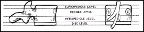
- Normal disc